

Trusted medical expertise for your family's health and well-being.
Established in November 1999, Sunway Medical Centre is an Australian Council on Healthcare Standards (ACHS) International and Malaysian Society for Quality in Health (MSQH) accredited private hospital. Located in the smart sustainable Sunway City, Sunway Medical Centre is surrounded by an ecosystem that will inspire a healthy, safe and interconnected society for generations to come. This includes the Sunway Resort Hotel & Spa, Sunway Pyramid mall, Sunway Lagoon theme park, Sunway University and Monash University, which are all within walking distance from the hospital.
Learn More About us
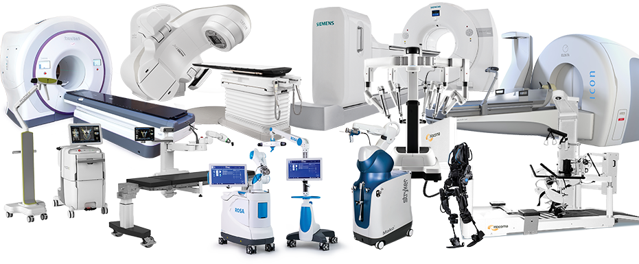
Your health is our priority. With comprehensive healthcare services, cutting-edge technology and facilities and our team of experts, we’re committed to delivering the highest standards of treatment and care for you and your loved ones.
Medical Procedures
Medical Specialities
Centres of Excellence
Licensed Beds
Critical Care Unit Beds
Operating Theatres
Catheterisation Labs
Haemodialysis Bays
Qualified Nurses
Accident & Emergency
Doctors with Practising Rights
Parking Spaces, Including Valet Parking
Radiology, Diagnostics, Laboratory, Blood Bank & Pharmacy Services
Children's Emergency
Our hospital welcomes patients from all over the world. To ensure a seamless and comfortable experience, our dedicated team offers international patients the support and guidance they need when seeking medical treatments at our hospital, including assistance with necessary arrangements.